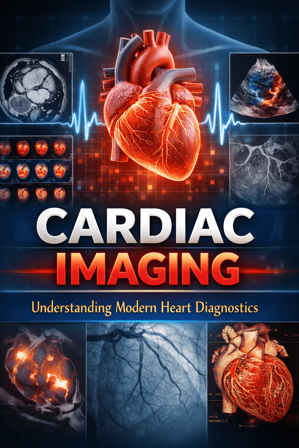
Cardiac Imaging: Understanding Modern Heart Diagnostics
Introduction
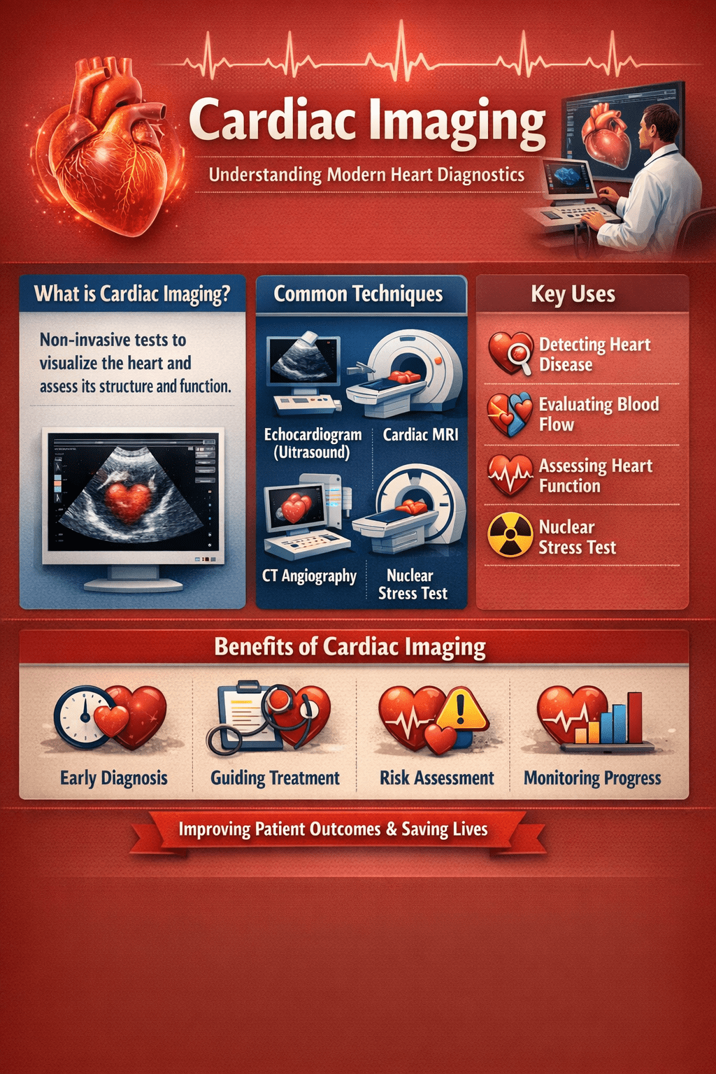
Cardiac Imaging is a medical tool that provides detailed pictures of the heart’s structure and function. It helps doctors assess heart chambers, valves, blood flow, and muscle performance. Common tests include echocardiograms, cardiac CT, cardiac MRI, and nuclear imaging. These tests are used to diagnose heart disease, evaluate symptoms such as chest pain or shortness of breath, and guide treatment decisions. As heart disease remains a leading cause of illness in the UK and USA, cardiac imaging has become central to early detection, accurate decision-making, and long-term heart care.
Cardiac Imaging also allows clinicians to detect subtle changes in the heart that might not yet cause noticeable symptoms, making it an essential tool for preventive care. By visualizing the heart in motion, doctors can monitor disease progression, evaluate the effectiveness of treatments, and plan interventions more accurately. Patients with risk factors such as high blood pressure, diabetes, or a family history of heart disease often benefit from these tests, which provide objective information to guide lifestyle adjustments or medical therapy. Ultimately, Cardiac Imaging gives both clinicians and patients a clearer understanding of heart health, supporting more informed, timely, and effective care decisions.
What Is Cardiac Imaging?
Cardiac imaging uses ultrasound, X-rays, magnetic fields, or radioactive tracers to visualize the heart’s chambers, valves, arteries, and blood flow without invasive procedures. These tests detect issues like coronary artery disease (CAD), heart failure, valve disorders, or plaque buildup early. Common applications include evaluating chest pain, shortness of breath, or risk factors such as family history and high blood pressure.
Cardiac Imaging: Understanding Modern Heart Diagnostics
Types of Tests
Echocardiograms (echo) provide real-time images of heart valves and ejection fraction using ultrasound, lasting 30-60 minutes with no radiation. Cardiac CT or coronary calcium scans quickly assess artery plaque and calcium scores, ideal for risk screening in 10 minutes. Cardiac MRI offers detailed tissue views without radiation, taking 60-90 minutes for complex cases like cardiomyopathy. Nuclear tests (PET/SPECT) track blood flow with tracers during stress, helping diagnose CAD.
| Test Type | Radiation | Cost Range (USA/UK) | Duration | Best For |
| Echo | No | $300-1,000 / £500-1,500 | 30-60 min | Valves, function |
| CT/CAC | Yes | $500-3,000 / £250-600 | 10 min | Arteries, plaque |
| MRI | No | $1,000-5,000 / Varies | 60-90 min | Tissue detail |
| Nuclear (PET/SPECT) | Yes | $600-5,000 / Higher | 30-60 min | Blood flow |
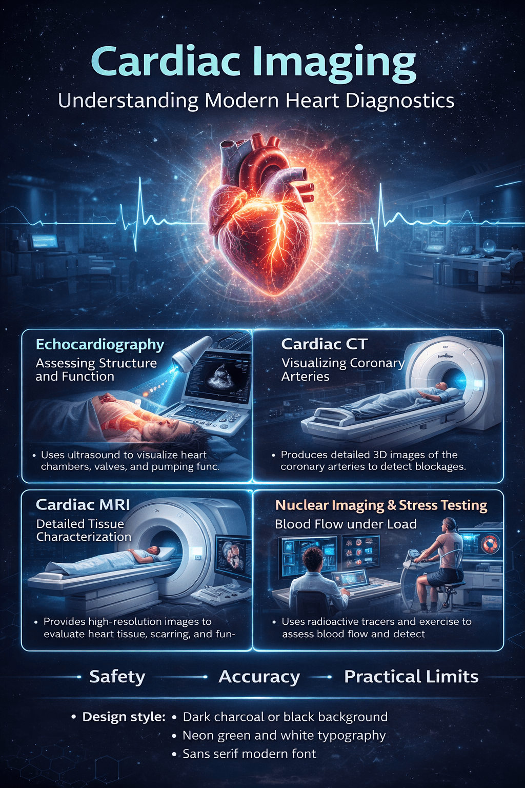
Echocardiography: Assessing Structure and Function
An echocardiogram uses ultrasound to show the heart in motion. It measures pumping strength, valve movement, and chamber size in real time.
This test matters because it quickly identifies common problems such as valve disease or reduced ejection fraction. In practice, it is often the first imaging test ordered for breathlessness or suspected heart failure.
Cardiac CT: Visualizing Coronary Arteries
Cardiac CT uses X-ray technology to create high-resolution images of the heart and coronary arteries. A coronary calcium scan detects plaque, while CT angiography shows narrowing or blockages.
These scans matter because they reveal coronary artery disease before symptoms become severe. They are frequently used for chest pain assessment or risk evaluation in people without known heart disease.
Cardiac MRI: Detailed Tissue Characterization
Cardiac MRI uses magnetic fields to produce detailed images of heart muscle and tissue composition. It can detect scarring, inflammation, and subtle structural changes.
This level of detail matters when diagnosing cardiomyopathy or assessing damage after a heart attack. In real-world care, MRI often answers questions other tests cannot.
Nuclear Imaging and Stress Testing: Blood Flow under Load
Nuclear cardiac imaging assesses how well blood reaches the heart muscle, often during physical or medication-induced stress. It highlights areas with reduced perfusion.
This matters because some heart problems appear only when the heart works harder. Stress imaging helps determine whether symptoms are caused by restricted blood flow.
Safety, Accuracy, and Practical Limits
Most cardiac imaging tests are safe and widely used. Some involve radiation or contrast agents, while others do not.
The key limitation is that no single test answers every question. Doctors balance accuracy, safety, cost, and patient factors when choosing the right approach.
Cardiac Imaging: Understanding Modern Heart Diagnostics
Conclusion
Cardiac imaging has reshaped how heart disease is detected and managed, shifting care from assumption to evidence. By matching the right test to the right clinical question, clinicians gain clarity while patients avoid unnecessary procedures. As technology advances, cardiac imaging will continue to refine diagnosis, personalize care, and support better long-term heart health. Those considering testing should use it as a tool for informed, measured decisions.
Medical Disclaimer: This guide provides general information, not medical advice. Consult a healthcare professional for personalized recommendations. Seek emergency care for severe chest pain or breathing issues.
Recommendation: How to Use Cardiac Imaging Wisely
If cardiac imaging is recommended, ask what question the test is meant to answer. Clarify whether it evaluates structure, blood flow, or risk.
Confirm preparation requirements, such as fasting or avoiding caffeine. Discuss potential follow-up steps if results are abnormal.
For people without symptoms, imaging should be guided by risk factors rather than curiosity alone. Used appropriately, cardiac imaging supports early detection and informed decision-making.
FAQs
What is cardiac imaging used for?
It is used to diagnose heart disease, assess symptoms, and guide treatment decisions.
Is cardiac imaging safe?
Yes. Most tests are noninvasive, with risks carefully managed.
How long do cardiac imaging tests take?
Anywhere from a few minutes to about 90 minutes, depending on the test.


